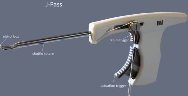
Kurt E.StollM.D.a, BenjaminHendyM.D.a, TregBrownM.D.b, NathanielCohenM.D.c, Thay Q.LeePh.D.d, SurenaNamdariM.D., M.Sc.a, PhilDavidsonM.D.e a Rothman Institute of Orthopaedics at Thomas Jefferson University Hospitals, Philadelphia, Pennsylvania, U.S.A. b The Orthopaedic Institute, Carbondale, Illinois, U.S.A. c OrthoNorCal, Los Gatos, California, U.S.A. d Orthopaedic Biomechanics Laboratory, Congress Medical Foundation, Pasadena, California, U.S.A. e Davidson Orthopedics, Salt Lake City, Utah, U.S.A. Received 11 October 2021, Accepted 24 December 2021, Available online 22 April 2022. Abstract Acromioclavicular (AC) joint injuries occur with a traumatic load to the lateral aspect of the shoulder and account for 9% of all shoulder girdle injuries. Rockwood classified AC joint injuries as type I though type VI, based on severity of injury, radiographic findings, and reducibility of the AC joint. Type I and II injuries are typically managed nonoperatively, whereas type IV, V, and VI are managed operatively to address the significant soft tissue disruption, persistent AC joint instability, and apical shoulder deformation. Treatment of type III injuries remain controversial. Several techniques have been described to treat AC joint injuries with no consensus for optimal treatment. “Anatomic” double-tunnel coracoclavicular ligament reconstruction is one currently popular technique to address AC joint injuries; however, clavicle and coracoid fractures are well-described complications of this technique. The objective of this technical report is to describe our preferred technique to address AC joint injuries. This technique involves using a looped braided polyester prosthetic band and low-profile buckle with allograft augmentation using a device to pass materials around the coracoid process. Technique Video Media player Download : Download video (62MB) Video 1. Patient with a Grade 4 AC dislocation in beach chair position. AC joint, coracoid, clavicle and planned clavicle incision is marked out. Posterior viewing portal is established and anterior rotator interval portal is used to debride around coracoid. Accessory anterior-inferior portal is established to see the base of the coracoid and resect tissue inferiorly. The open incision over the clavicle is made. This is a chronic case, so the incision is made at lateral edge of the clavicle. Clavipectoral fascia is released to establish pathway under the clavicle. The J-Pass device is loaded with a passing suture. An animated video is shown to explain how the J-Pass device is used to pass suture around the coracoid. The J-Pass is then used to pass a passing suture from medial to lateral around the coracoid. The suture is then passed under the clavicle. The M-Fix and graft are passed under the coracoid and clavicle. The AC joint is reduced, and the M-Fix is provisionally fixed. Reduction is confirmed with arthroscopy. The M-Fix is locked, and the tail is cut. The graft is sutured to itself. Introduction Injuries to the acromioclavicular (AC) joint are common in the active adult population and most often occur from a fall onto the lateral shoulder. This results in direct force onto the superior aspect of the acromion process, resulting in injury. The Rockwood classification of AC joint injuries helps guide management, with type I and II injuries typically managed conservatively and type IV to VI with surgical intervention. Treatment for type III injuries remains controversial, and choice of treatment depends upon an array of physician and patient related factors. There is no gold standard for the surgical management of AC joint separations with more than 150 surgical techniques described in the literature.1 Surgical techniques include open and arthroscopic approaches to reconstruct the coracoclavicular (CC) ligaments and/or AC ligaments with use of tendon autograft or allograft, temporary joint stabilization with hardware, distal clavicle excision, and CC space-stabilizing procedures using suture loop, screw, button, suture anchors, or similar devices. However, surgical treatment of AC joint injuries has been fraught with high rates of failure and complications. Reports show up to 80% of patients have loss of radiographic reduction and 20-30% of patients managed surgically undergo reoperation due to complications.2,3 Anatomic double-tunnel CC ligament reconstruction is one of the most common techniques to address AC joint injuries, as described by Mazzocca et al.4 Other techniques use single-drill tunnels into the clavicle and coracoid.5 Inherent in these techniques are two bone tunnels in the distal clavicle, which have been shown in biomechanical studies to weaken the clavicle, and there have been several case reports of clavicle fractures.6,7 Other techniques require drilling into the coracoid, which also raises concern for coracoid fractures.8 In an effort to mitigate the morbidity of bone tunnels in the distal clavicle and lessen loss of reduction, we describe a tunnel-free technique involving the use of a broad polyester suture band (4 mm or 7 mm) with a preattached metal buckle and allograft augmentation (see Table 1 for advantage and disadvantages). Table 1. Advantages and Disadvantages Advantages Disadvantages 1. No bone tunnels are created in the clavicle or coracoid; therefore, the bone is not weakened. 1. More extensive dissection is needed around the clavicle in order to allow passage of the graft and M-fix. 2. M-Fix has a looped woven prosthetic band configuration. 2. In order to successfully pass the graft and M-fix around the coracoid, adequate soft tissue dissection must be done, putting neurovascular structures at risk. 3. The M-Fix can be used as provisional fixation prior to final locking, which enables assessment of reduction on fluoroscopy. 3. There is a risk of hardware prominence on the clavicle. Surgical Technique For details on the surgical technique, see Video 1 and Tables 2 and 3. Table 2. Steps Step 1: Patient Positioning • Beach chair • Drape to allow a wide access to the posterior and medial shoulder. • Ensure access for fluoroscopy. Step 2: Diagnostic Glenohumeral Arthroscopy • Standard posterior viewing portal • Anterior portal through rotator interval • Perform diagnostic arthroscopy. Step 3: Coracoid Process Exposure • Work laterally to medially. • Debride lateral and inferior aspect of coracoid process. • An accessory anterior-inferior portal helps with visualization. Step 4: Mini-Open Exposure of Clavicle • Incision is 4 cm medial to the AC joint. • Expose clavicle anteriorly and posteriorly • Clear soft tissue from the clavicle at the level of the coracoid process. Step 5: Subcoracoid Passage Preparation • Reinsert arthroscope through posterior portal. • Ensure there is a clear path between clavicle and coracoid process. • May use a blunt obturator through clavicle incision to clear out soft tissue. Step 6: Subcoracoid Passage of Shuttling Suture

Orthopaedic Institute of Southern IL - Herrin (Main Campus)
Orthopaedic Institute of Southern IL - Herrin (Main Campus)
Deaconess Crossroads Hospital - Mt. Vernon
OISI - Carbondale
SIU Student Health Center - Carbondale
Harrisburg Medical Center - Harrisburg
SIH - Herrin Hospital
Deaconess Medical Center IL - Marion
SIH - Memorial Hospital of Carbondale
Southern Illinois Orthopedic Center (SIOC) - Herrin (Main Campus)
Marshall Browning Hospital - Du Quoin
Franklin Hospital - Benton
Fairfield Memorial Hospital - Fairfield
618-997-6800